High-Definition Mapping of Four Spatially Distinct Neutralizing Epitope Clusters on RiVax, a Candidate Ricin Toxin Subunit Vaccine

Abstract
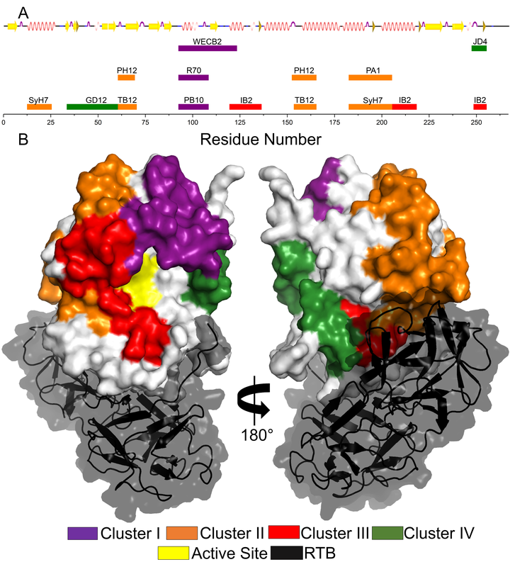
RiVax is a promising recombinant ricin toxin A subunit (RTA) vaccine antigen that has been shown to be safe and immunogenic in humans and effective at protecting rhesus macaques against lethal-dose aerosolized toxin exposure. We previously used a panel of RTA-specific monoclonal antibodies (MAbs) to demonstrate, by competition enzyme-linked immunosorbent assay (ELISA), that RiVax elicits similar serum antibody profiles in humans and macaques. However, the MAb binding sites on RiVax have yet to be defined. In this study, we employed hydrogen exchange-mass spectrometry (HX-MS) to localize the epitopes on RiVax recognized by nine toxin-neutralizing MAbs and one nonneutralizing MAb. Based on strong protection from hydrogen exchange, the nine MAbs grouped into four spatially distinct epitope clusters (namely, clusters I to IV). Cluster I MAbs protected RiVax’s α-helix B (residues 94 to 107), a protruding immunodominant secondary structure element known to be a target of potent toxin-neutralizing antibodies. Cluster II consisted of two subclusters located on the “back side” (relative to the active site pocket) of RiVax. One subcluster involved α-helix A (residues 14 to 24) and α-helices F-G (residues 184 to 207); the other encompassed β-strand d (residues 62 to 69) and parts of α-helices D-E (154 to 164) and the intervening loop. Cluster III involved α-helices C and G on the front side of RiVax, while cluster IV formed a sash from the front to back of RiVax, spanning strands b, c, and d (residues 35 to 59). Having a high-resolution B cell epitope map of RiVax will enable the development and optimization of competitive serum profiling assays to examine vaccine-induced antibody responses across species.